

Calcific Tendonitis
What is it?
Calcific tendonitis of the shoulder happens when calcium deposits form on the tendons of your shoulder. The tissues around the deposit can become inflamed, causing a great deal of shoulder pain.
Which part of the shoulder is affected?
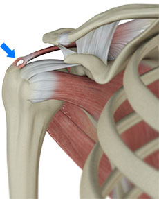
Calcific tendonitis occurs in the tendons (tendons attach muscles to bones) of the rotator cuff. The rotator cuff is actually made up of several tendons that connect the muscles around your shoulder to the humerus (the larger bone of the upper arm). Calcium deposits usually form on the tendon in the rotator cuff called the supraspinatus tendon. Why it occurs is not clear. It doesn't seem to be related to degeneration and it is more likely to cause shoulder pain than degenerative calcification, in which calcium forms due to a "wear and tear" process.
Doctors think of calcific tendonitis, or reactive calcification, in three stages:
In the pre-calcific stage, the tendon changes in ways that make calcium deposits more likely to form. In the calcific stage, calcium crystals are deposited in the tendons. Then they begin to disappear as the body simply reabsorbs the calcium deposits. Ironically, it is during this stage that pain is most likely to occur. In the post-calcific stage, the body heals the tendon, and the tendon is remodeled with new tissue. No one knows what triggers the body to reabsorb the deposits. But once this occurs and the tissue begins to be remodeled, the pain usually decreases or goes away altogether.
Why did I develop calcific tendonitis?
No one really knows what causes calcific tendonitis. This type of problem occurs in younger patients and seems to go away by itself in many cases.
What are the symptoms?
While the calcium is being deposited, you may feel only mild to moderate pain, or even no pain at all. For some unknown reason, calcific tendonitis becomes very painful when the deposits are being resorbed. The pain and stiffness of calcific tendonitis can cause you to lose motion in your shoulder. Lifting your arm may become painful. At its most severe, the pain may interfere with your sleep. Symptoms improve once the calcium is resorbed.
Some people only experience a single episode of pain, some get two or three, and some people get regular flare-ups of symptoms over many years. Also, in between the painful episodes, some people may have no symptoms at all while others may have a constant pain.
How is it diagnosed?
Your doctor will take a detailed medical history and perform a thorough physical examination of your shoulder. The pain of calcific tendonitis can be confused with other conditions that cause shoulder pain. An X-ray is usually necessary to confirm the presence of calcium deposits. You may need to get several X-rays over time. This will help your doctor keep track of the changes in the amount of calcification. By following the changes in the calcium deposits, your doctor can determine whether the condition will heal by itself or perhaps require surgery. Ultrasound scans are also very good at determining the extent of calcific deposits.
What is the treatment?
The main aim of treatment is to control the pain and maintain the function of the shoulder. Unfortunately, nothing has been shown to affect the natural course of the condition itself.
Non-surgical treatment:
Initial treatment is likely to be rest and anti-inflammatory medication, such as ibuprofen. The anti-inflammatory medicine is used mainly to control pain.
Your doctor may suggest a cortisone injection if your pain stays severe even after trying other nonsurgical treatments. Cortisone is a very powerful steroid. Cortisone can be very effective at temporarily easing inflammation and swelling.
During the time when the calcium deposits are being resorbed, the pain can be excruciating and require urgent treatment. An effective treatment during this phase is to have the calcific deposits needled under ultrasound guidance, combined with a local anaesthetic and steroid injection.
Physiotherapy is also focused on easing your pain and inflammation. Treatments may include heat or ice.
Shock wave therapy is a newer form of nonsurgical treatment. It uses a machine to generate shock wave pulses to the sore area. Patients generally receive the treatment once each week for up to three weeks. The impulses are thought to help break up the deposit so the body can more easily absorb it. Recent studies indicate that this form of treatment can help ease pain and reduce the size of the deposit.
Surgical treatment:
Surgery is reserved for cases when all other methods have failed to control the symptoms. A special TV camera called an arthroscope is inserted into the shoulder joint through a small incision in the skin. Other small incisions allow the surgeon to insert small surgical instruments into the joint as well. The surgeon uses the arthroscope to locate the calcium deposit in the rotator cuff tendon. Once the deposit is found, the surgeon uses the small instruments to resect (remove) the calcium deposits and rinse the area. This is usually combined with a subacromial decompression, which increases the space available for the affected tendon.